KINGDOMTOTO: SITUS TOTO DENGAN LIVE RESULT TERCEPAT TERUPDATE
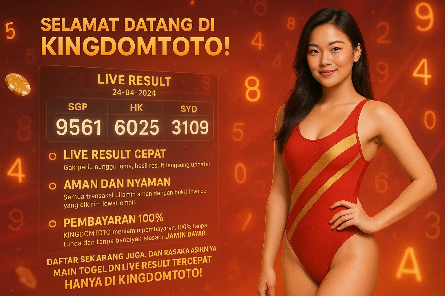
Selamat datang di KINGDOMTOTO! Nikmati serunya live result dan live draw, terkoordinasi langsung dengan situs resmi. Hasil selalu 100% akurat dan terbaru untuk pasaran SGP, HK, dan SDY.
Nikmati Semua Hanya di KINGDOMTOTO:
- Live Result Cepat: Hasil langsung terupdate tanpa delay.
- Aman & Terpercaya: Transaksi dijamin, lengkap dengan invoice via email.
- Pembayaran 100%: Kami bayar penuh, tanpa tunda dan alasan.
📌 Latest Posts
Loading posts…
Memuat posting…
Live Result Terupdate
Loading live result…
Sedang Menyusun Data Result Terbaru…